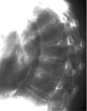
Bilateral hip DXA scan image from a 59-year-old post-menopausal woman.
Price: $ 19.00
4.7(365)
Download scientific diagram | Bilateral hip DXA scan image from a 59-year-old post-menopausal woman. The dominant arm did not match, but dominant leg did. The T-score for the lumbar spine was normal. If the patient had only had her left hip examined in accordance with the dominant arm, the conclusion would have been normal bone mineral density (BMD). Having both hips examined instead led to the conclusion of low bone density (LBD). from publication: Dual-energy X-ray Absorptiometry of Both Hips Helps Appropriate Diagnosis of Low Bone Mineral Density and Osteoporosis | Controversy still remains regarding the use of bilateral hip scanning when bone mineral density (BMD) is measured, and bilateral hip scanning is not mandatory in international guidelines for screening of osteoporosis. BMD of both hips and the lumbar spine was analyzed in 133 | Hip, Dual-Energy X-ray Absorptiometry and Bone Mineral Density | ResearchGate, the professional network for scientists.

Movement during DXA imaging can have a deleterious effect on image
Anatomical context of focal thinning in women with femoral neck

Best Practice Recommendations for DXA Scans and Reports

Evolutionary roots of the risk of hip fracture in humans

Composite indices of femoral neck strength predicts the collapse of steroid-associated osteonecrosis of the femoral head: a retrospective study, BMC Musculoskeletal Disorders
Osteoporosis Workup: Approach Considerations, Laboratory Studies, Biochemical Markers of Bone Turnover

Osteoporosis Imaging: State of the Art and Advanced Imaging

Bone Metabolism

JCM, Free Full-Text
Cureus, Osteoporosis in a 60-Year-Old Male With a History of Chronic Myeloid Leukemia Treated With Imatinib Mesylate

Bone Mineral Densitometry Reporting: Pearls and Pitfalls - Patrick Martineau, Sarah L. Morgan, William D. Leslie, 2021

Bilateral hip DXA scan image from a 59-year-old post-menopausal woman.

Quality in dual-energy X-ray absorptiometry scans - ScienceDirect

Bone Mineral Densitometry Reporting: Pearls and Pitfalls - Patrick Martineau, Sarah L. Morgan, William D. Leslie, 2021